![]() Figure 6 of
Gilliland, Mol Vis 2001;
7:120-130.
Figure 6 of
Gilliland, Mol Vis 2001;
7:120-130.
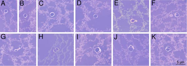
Figure 6. Gallery of light micrographs
These MLBs from the fetal nucleus display varying geometrical patterns. MLBs in A, B, C, and D are circular. The MLB in E shows a small protrusion. MLBs in F through K display doublet and even triplet structures.