![]() Figure 3 of
Gilliland, Mol Vis 2001;
7:120-130.
Figure 3 of
Gilliland, Mol Vis 2001;
7:120-130.
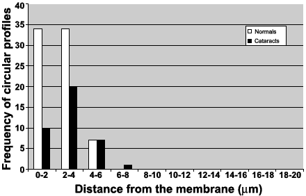
Figure 3. Distance mapping of profiles from the membrane
Quantitative measurements were made from four normal lenses and four cataracts. Circular profiles were counted in random areas (7000 mm2) of embryonic and fetal nuclear regions, and the distance between each profile and the cell membrane was measured. Results demonstrate that the normal lens contains more circular profiles than the cataract, although the difference is not statistically significant. These profiles are predominantly located within 4 mm of the cell membrane.