![]() Figure 6 of
Gendron, Mol Vis 2001;
7:107-113.
Figure 6 of
Gendron, Mol Vis 2001;
7:107-113.
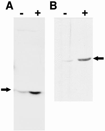
Figure 6. Regulation of gene expression in MK/T-1 cells by TGF-b2 treatment
Induction of activating phosphorylation of p38-MAPK (A), and induction of a-smooth muscle actin (SMA-a, B) by TGF-b. Western blot analysis with a rabbit anti-phospho-p38-MAPK antibody (A) and a mouse anti-SMA-a (B) of extracts (50 mg of protein) of MK/T-1 cells unstimulated (-) and stimulated (+) with TGF-b2. MK/T-1 cells treated in culture for 1 h with TGF-b2 showed a significant induction of phospho-p38-MAPK (plus lane in A), while the untreated MK/T-1 cells expressed very low levels of phospho-p38-MAPK at 1 h (minus lane in A). MK/T-1 cells treated in culture for 48 h with TGF-b2 showed a significant induction of SMA-a (plus lane in B), while the untreated MK/T-1 cells expressed very low levels of SMA-a at 48 h (minus lane in B). Arrows indicate positions of phospho-p38-MAPK and a-smooth muscle actin bands in A and B.