![]() Figure 2 of
Wu, Mol Vis 2001;
7:101-106.
Figure 2 of
Wu, Mol Vis 2001;
7:101-106.
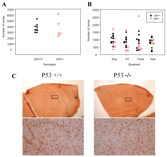
Figure 2. p53 independent cone cell degeneration
Twenty one 0.25 mm x 0.25 mm boxes per quadrant (eighty four per eye) were used to count PNA positive cones in whole mount retinas. Retinas from three month old mice of the indicated genotypes were stained and total counts (A), or the number of cones in each quadrant (B) were plotted. Nine p53+/+ and six p53-/- eyes were analyzed. Typical PNA stained whole mounts are shown in C. Individual PNA positive cone cells are visible in the lower panels, which represent magnified views of the boxed region in the upper panels. Similar numbers of cones were observed in the presence or absence of p53