![]() Figure 4 of
Wang, Mol Vis 2001;
7:89-94.
Figure 4 of
Wang, Mol Vis 2001;
7:89-94.
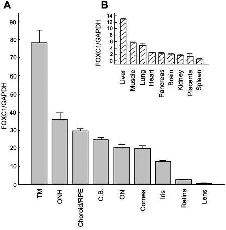
Figure 4. FOXC1 expression in human tissues
RNA from ocular tissue dissected from four different donor eyes was pooled and reverse transcribed with random primers for quantification by real-time PCR. A: FOXC1 expression (presented in order of relative abundance) in trabecular meshwork (TM), optic nerve head (ONH), choroid/RPE, ciliary body, optic nerve (ON), cornea, iris, retina, and lens was normalized to GAPDH expression levels in the same cDNA samples. Each bar represents the mean ± the standard deviation of 3 replicate analyses. B (inset): FOXC1 expression in non-ocular human tissues by real-time PCR normalized to GAPDH expression levels.