![]() Figure 2 of
Lyubarsky, Mol Vis 2001;
7:71-78.
Figure 2 of
Lyubarsky, Mol Vis 2001;
7:71-78.
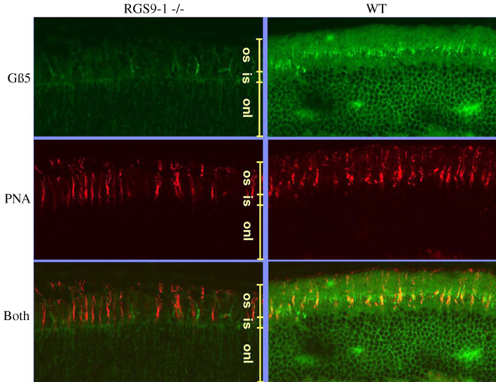
Figure 2. Immunofluorescence staining of Gb5 and cones in the mouse retina
Left column: section from retina of an RGS9-1 -/- mouse. Right column: sections from wildtype (C57BL/6) mouse. "Gb5", staining with fluorescein-isothiocyanate (FITC) conjugated to secondary antibody, with primary against Gb5, as described in Methods. "PNA", staining with rhodamine-conjugated peanut agglutinin (red), which binds to cones. The two lowermost panels ("Both") were prepared by digitally summing the fluorescein and rhodamine images.