![]() Figure 7 of
Boyle, Mol Vis 2000;
6:63-71.
Figure 7 of
Boyle, Mol Vis 2000;
6:63-71.
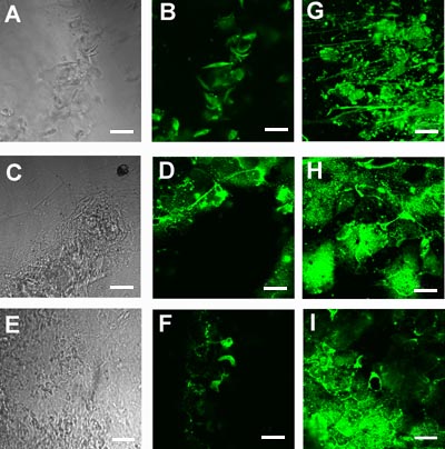
Figure 7. Immunolocalization of markers for lens cell differentiation
Representative individual confocal optical sections of the immunolocalization of MIP 26 (B, G), b-crystallins (D, H), and g-crystallins (F, I) in cultures 4-7 days after loading with a-crystallins. A, C, and E are corresponding DIC images for images in B, D, and F. MIP 26, b- and g-crystallins were localized to cells within large aggregates (B, D, and F) and in cells present in stratified cell layers (G, H, and I). Images B and F were taken from an optical plane distal from the dish bottom, while images D, G, H, and I were from optical planes more proximal to the dish bottom.