![]() Figure 6 of
Boyle, Mol Vis 2000;
6:63-71.
Figure 6 of
Boyle, Mol Vis 2000;
6:63-71.
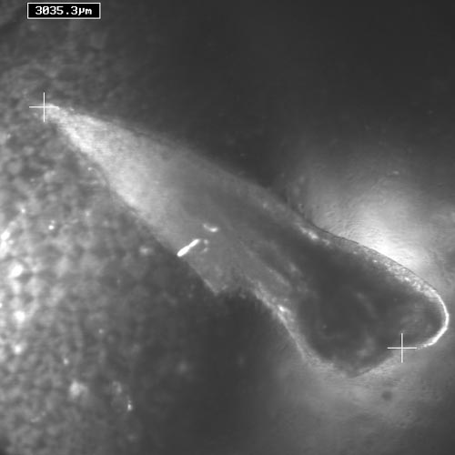
Figure 6. A primary bovine lens epithelial culture 4 days after loading with aB-crystallin
Representative DIC image of a primary lens epithelial culture 4 days after loading cells with aB-crystallin. In the one exception out of 8 cultures that were followed, a large multilayered, multicellular structure was observed 4 days after loading primary bovine lens epithelial cells with aB-crystallin. The number in the upper left corner is the distance between the two white plus marks on the image. This structure measured approximately 3 mm in length by 0.8 mm in width.