![]() Figure 5 of
Boyle, Mol Vis 2000;
6:63-71.
Figure 5 of
Boyle, Mol Vis 2000;
6:63-71.
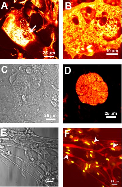
Figure 5. Primary bovine lens epithelial cultures loaded with Texas Red labeled a-crystallins
Representative DIC images (C, E) with corresponding individual confocal optical sections, approximately 0.5 mm thick, (D, F) of cells loaded with Texas Red labeled a-crystallins and individual confocal optical sections (A, B) of cells loaded with Texas Red labeled a-crystallins at various times postloading. Images A and B were taken within the first h after loading with protein and showed a-crystallins localized to cytoplasmic structures (A, B) and within the nucleus (A, arrow). Images C and D were taken approximately 4 h after loading with protein. At this time, multilayered aggregated masses of cells were observed (C) that contained a-crystallins throughout the cytoplasm of cells within the aggregate (D). By 4 days postloading, elongated cells similar in dimensions to elongating fiber cells in the bow region of the lens were observed (E). These cells contained Texas Red labeled a-crystallins localized diffusely throughout the cytoplasm that was of weak fluorescence intensity compared to 1-4 h postloading (F). Images E and F were taken from the edge of a large aggregate of cells. Cells in image F had been fixed, permeablized, and stained with SYTOX GREEN (Molecular Probes) to visualize cell nuclei. Many of the nuclei appear condensed and elongated (arrowheads), similar to nuclei seen in lens fiber cells in the bow region of the lens.