![]() Figure 4 of
Boyle, Mol Vis 2000;
6:63-71.
Figure 4 of
Boyle, Mol Vis 2000;
6:63-71.
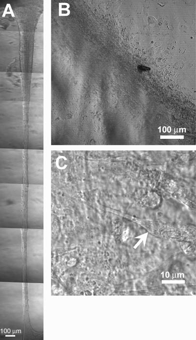
Figure 4. Primary bovine lens epithelial cultures 4-7 days after loading with a-crystallins and WSF
Representative DIC images of primary lens epithelial cultures, 4-7 days after loading cells with a-crystallins (A) or WSF (B, C), showing large multilayered, multicellular masses of cells. Some of the cells within these masses are similar in dimensions to elongating fiber cells within the bow region of the lens (C, arrow).