![]() Figure 3 of
Boyle, Mol Vis 2000;
6:63-71.
Figure 3 of
Boyle, Mol Vis 2000;
6:63-71.
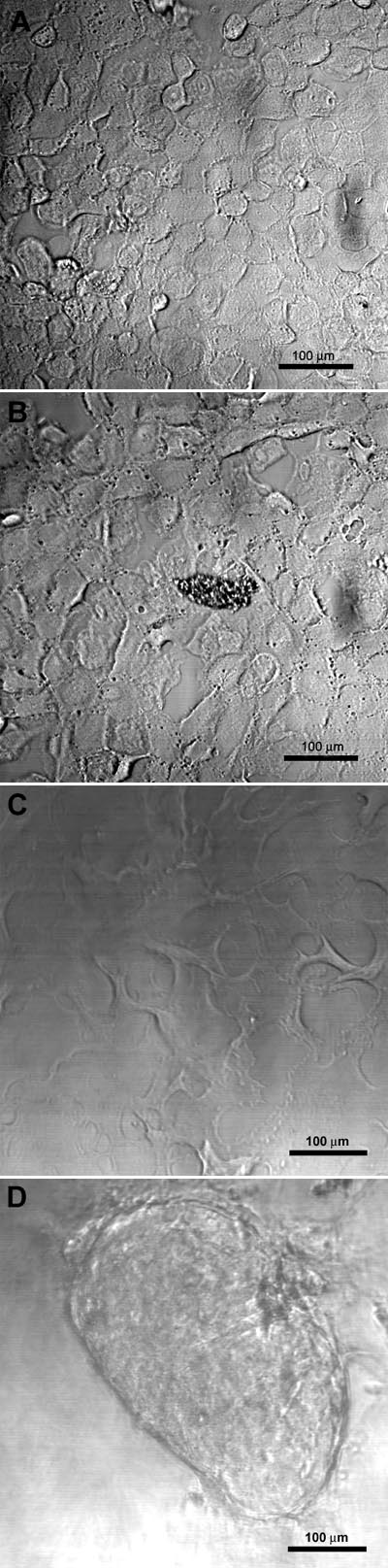
Figure 3. Primary bovine lens epithelial cultures 0-3 h after loading with aA- or aB-crystallins
A. Primary bovine lens epithelial cells prior to loading with crystallins. B. A representative DIC image of a culture 3 h post loading with aA-crystallin. C. A representative DIC image of a culture 3 h post loading with aB-crystallin in an area not containing a lentoid body. D. A representative DIC image of the same culture in (C) with a lentoid body.