![]() Figure 1 of
Boyle, Mol Vis 2000;
6:63-71.
Figure 1 of
Boyle, Mol Vis 2000;
6:63-71.
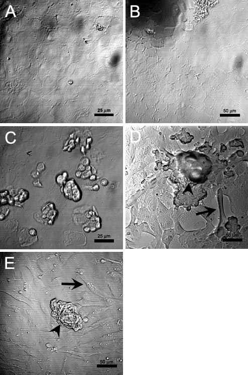
Figure 1. Primary bovine lens epithelial cultures 1-2 h after protein loading
Representative differential interference contrast (DIC) images of primary lens epithelial cultures, 1-2 h after loading with: (A) sham procedure, (B) BSA, (C) BSA, (D) WSF, and (E) a-crystallins. Sham (A) and BSA loaded (B) cells show no changes in morphology from pretreated cultures, which were a monolayer of epithelial cells. An increased number of cells not adherent to the dish are observed in some areas of a given culture (C) after osmotic lysis of pinosomes. Aggregation (arrowheads) and elongation (arrows) of epithelial cells are apparent in WSF- (D) and a-crystallins loaded cells (E).