![]() Figure 5 of
Gross, Mol Vis 2000;
6:51-62.
Figure 5 of
Gross, Mol Vis 2000;
6:51-62.
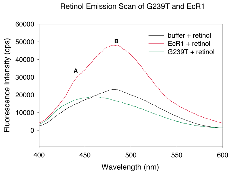
Figure 5. Retinol emission scan of G239T and EcR1
Emission scans from 400 to 600 nm of 1 mM wild type EcR1, G239T or protein-free buffer plus 10 mM all-trans-retinol were carried out with excitation set at 330 nm. The scan of retinol-EcR1 (red line) revealed a maximum at 479 nm (label "B") and a small shoulder at 440-450 nm (label "A"). G239T (green line) complexed with retinol exhibited a maximum at about 450 nm, and the peak height was much lower. Retinol in buffer without protein is represented by the black line. The comparatively high concentration of free retinol (~9 mM) partly masks the small fluorescence enhancement contributed by the ~1 mM G239T-retinol complex indicated by the green line. One explanation of the shift in emission maxima is that the mutation causes a reduction in fluorescence intensity and shifts the maximum to 450 nm within a single ligand binding site. Another possible explanation is that a highly fluorescent site with a maximum at 479 nm is lost, making more apparent a second site (which contributes the shoulder at 450 nm) recognizable as a peak with a maximum at 450 nm in G239T.