![]() Figure 4 of
Gross, Mol Vis 2000;
6:51-62.
Figure 4 of
Gross, Mol Vis 2000;
6:51-62.
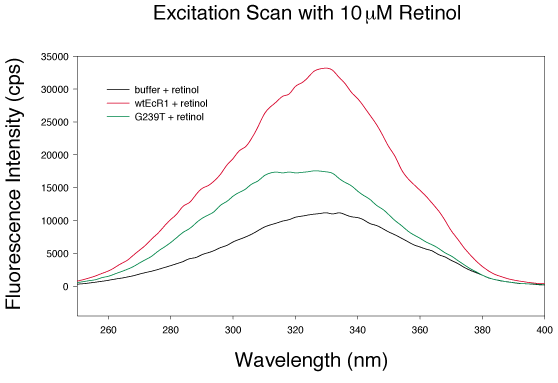
Figure 4. Retinol excitation scan of G239T and EcR1
An excitation scan from 250 to 400 nm of 1 mM wild type EcR1, G239T, or buffer plus 10 mM retinol was carried out (emission set to 479 nm). The excitation maxima for all samples was 336 nm. The red line and squares represents EcR1, the green line and triangles represents G239T, and the black line and circles represent buffer lacking protein.