![]() Figure 1 of
Gross, Mol Vis 2000;
6:51-62.
Figure 1 of
Gross, Mol Vis 2000;
6:51-62.
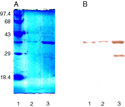
Figure 1. SDS PAGE and monoclonal antibody detection of G239T and EcR1
EcR1 and G239T were grown and purified as described [12]. The samples were run on two 10% SDS polyacrylamide gels [34]. In lane 1, EcR1 was mixed with molecular weight markers and the sizes of the markers are shown on the left. In lane 2 purified G239T was loaded. In Lane 3, about ten times the amount of G239T was loaded. (A) shows a Coomassie Brilliant Blue stained gel. (B) shows an immunoblot stained with the monoclonal antibody H3B5 [35]. The results showed that G239T and EcR1 migrated with the same mobility and gave an estimated mass of 36,400 Da. This is in close agreement with the calculated molecular mass of 34,500.4 Da. Both EcR1 and G239T showed strong reactivity with a monoclonal antibody specific for Repeats 1 and 2 of human IRBP [35]. The G239T preparations were >90% pure as indicated by the lack of other detectable bands on the protein stained gel. However, an apparently degraded product of G239T, with an estimated molecular mass of 27,500 Da was detected by immunostaining in Lane 3 of B. This band is virtually undetectable in the protein-stained gel.