![]() Figure 5 of
Leung, Mol Vis 2000;
6:15-23.
Figure 5 of
Leung, Mol Vis 2000;
6:15-23.
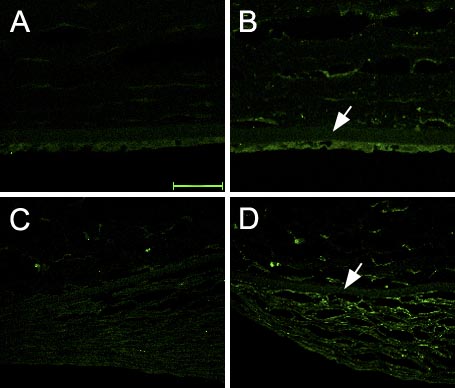
Figure 5. Immunofluorescent staining of the long splice-variant of type XII collagen
Normal cornea stained with anti-type XII collagen antibody (long variant form; B) and negative control performed using FITC-conjugated secondary antibodies made in different species (chick) from the mouse primary antibody (A). RCFM-containing cornea stained with anti-type XII collagen antibody (long variant form; D) and negative control performed using FITC-conjugated secondary antibodies made in different species (chick) from the mouse primary antibody (C). Bar is 50 mm. Arrow indicates the position of Descemet's membrane.