![]() Figure 4 of
Leung, Mol Vis 2000;
6:15-23.
Figure 4 of
Leung, Mol Vis 2000;
6:15-23.
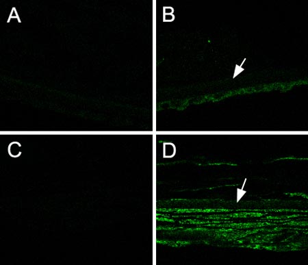
Figure 4. Immunofluorescent staining of type III collagen
Normal cornea stained with anti-type III collagen antibody (B) and negative control (A). RCFM-containing cornea stained with anti-type III collagen antibody (D) and negative control (C). Arrow indicates the position of Descemet's membrane.