![]() Figure 1 of
Grimm, Mol Vis 2000;
6:252-260.
Figure 1 of
Grimm, Mol Vis 2000;
6:252-260.
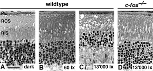
Figure 1. Photoreceptor apoptosis induced by high levels of white light
Light microscopic analysis of sections of central inferior retinal tissues of wildtype (129SV/Bl6) mice (A through C) and of c-fos-/- mice (D) before (A) and at 48 h after exposure to 13,000 lux (C, D) or to 60 lux (B) of white fluorescent light for 2 h. Scale bar: 25 mm. PE: pigment epithelium; ROS: rod outer segment; RIS: rod inner segment; ONL: outer nuclear layer.