![]() Figure 4 of
Yang, Mol Vis 2000;
6:237-242.
Figure 4 of
Yang, Mol Vis 2000;
6:237-242.
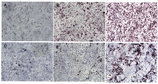
Figure 4. Immunohistochemical staining of Lentivirus-transduced ARPE-19 and COS-7 cells
ARPE-19 (A-C) and COS-7 cells (D-F) were transduced with the following different concentrations of recombinant Lentivirus: (A and D) 0.1 TU/cell; (B and E) 1 TU/cell; and (C and F) 10 TU/cell. After 2 days of incubation with Lentivirus, human RGR opsin was detected by immunohistochemical staining with the DE7 antibody [11]. Immunoreactivity was developed with the Vector VIP substrate, and the cells were counterstained with Mayer's hematoxylin.