![]() Figure 7 of
Choi, Mol Vis 2000;
6:222-231.
Figure 7 of
Choi, Mol Vis 2000;
6:222-231.
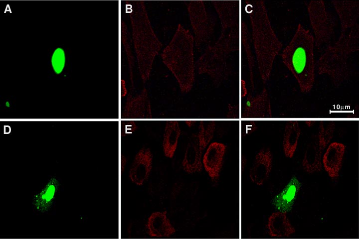
Figure 7. Expression of high affinity receptors of FGF
Cells were transfected with pEGFP-18-kDa FGF-2 and further incubated for 24 h (A, B, and C) or for 48 h (D, E, and F). At the end of each incubation, cells were fixed, permeabilized, and stained with monoclonal mouse anti-FGF-R-1 antibody (B and E). The fluorescent signal and Texas red signals were superimposed in C and F. Data are representative of three experiments. The scale bar represents 10 mm.