![]() Figure 4 of
Choi, Mol Vis 2000;
6:222-231.
Figure 4 of
Choi, Mol Vis 2000;
6:222-231.
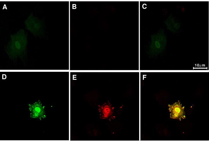
Figure 4. Subcellular localization of GFP/18-kDa FGF-2 48 h after transfection
Cells were transfected with either pEGFP-18-kDa FGF-2 (D, E, and F) or pEGFP-C3 (A, B, and C). After 48 h incubation, cells were fixed, permeabilized, and stained with anti-FGF-2 antibodies (B and E). The fluorescent signal and Texas red signals were superimposed in C and F. Data are representative of five experiments. The scale bar represents 10 mm.