![]() Figure 2 of
Choi, Mol Vis 2000;
6:222-231.
Figure 2 of
Choi, Mol Vis 2000;
6:222-231.
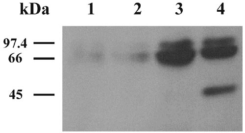
Figure 2. Immunoblotting analysis of expressed 18 kDa FGF-2
Cell lysates (30 mg) prepared from transfected CEC were separated on a 12.5% SDS-polyacrylamide gel under reducing conditions and transferred to PVDF membrane followed by immunoblotting with anti-FGF-2 antibody. Lane 1; CEC alone. Lane 2; CEC transfected with pEGFP-C3. Lane 3; CEC transfected with pEGFP-18-kDa FGF-2/R. Lane 4; CEC transfected with pEGFP-18-kDa FGF-2. Data are representative of three experiments.