![]() Figure 5 of
John, Mol Vis 2000;
6:204-215.
Figure 5 of
John, Mol Vis 2000;
6:204-215.
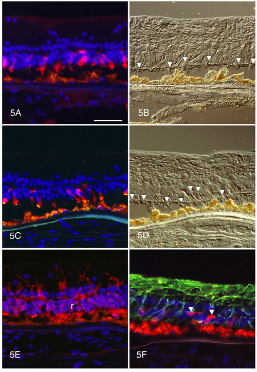
Figure 5. Immunocytochemistry of retina with G-106-R RHO mutation
A. All remaining cones are strongly positive with 7G6. Their outer segments are short to absent. B. The cones (arrowheads) are documented by DIC microscopy in the same field shown in Figure 5A. C. The shortened cone outer segments are positive for red/green cone opsin (gold). The cone cytoplasm is 7G6-positive (red). D. Nomarski image of field in Figure 5C illustrates the cones (arrowheads). E. As revealed by anti-rhodopsin labeling, the rods have tiny or absent outer segments and rhodopsin (red) is localized to the cell bodies (r) and neurites. F. Hypertrophied Müller processes are strongly positive for GFAP (green), and the few remaining cones (arrowheads) are well labeled with 7G6 (red).
Autofluorescent lipofuscin is present in the retinal pigment epithelium at the bottom of the panels. The scale bar represents 50 mm.