![]() Figure 4 of
John, Mol Vis 2000;
6:204-215.
Figure 4 of
John, Mol Vis 2000;
6:204-215.
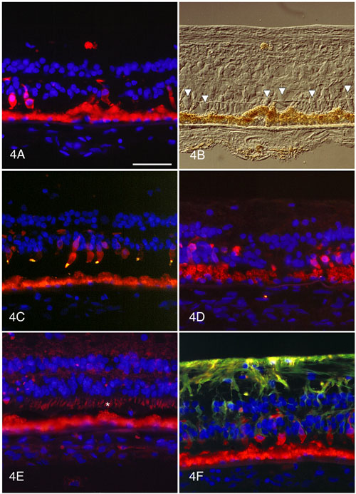
Figure 4. Immunocytochemistry of retina with P-23-H RHO mutation
A. Most remaining cones are well labeled with 7G6. B. DIC microscopy of same field as Figure 4A. The cones are indicated by the arrowheads. C. Some cones are well labeled with 7G6 (red) and most cone outer segments are positive (gold) for red/green cone opsin. D. IRBP immunolabeling (green) is absent from the interphotoreceptor matrix. The cones are 7G6-positive (red). The retinal pigment epithelium cells are enlarged and engorged with autofluorescent (red) lipofuscin granules. E. Cytochrome C oxidase labeling is weak in the shortened photoreceptor inner segments (*). F. GFAP labeling is strong in the hypertrophied Müller cell processes. Some cones, whose outer segments are tiny, are 7G6-positive (red).
Autofluorescent lipofuscin is present in the retinal pigment epithelium at the bottom of the panels. The scale bar represents 50 mm.