![]() Figure 3 of
John, Mol Vis 2000;
6:204-215.
Figure 3 of
John, Mol Vis 2000;
6:204-215.
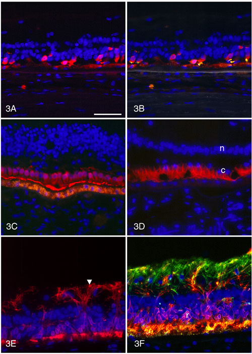
Figure 3. Immunocytochemistry of retina with Q-64-ter RHO mutation
A. All remaining cones are strongly labeled with 7G6. Their outer segments are very short. B. The shortened cone outer segments (gold) are positive for red/green cone opsin and 7G6 (red). C. The macular cones are reduced to a single layer of cell bodies that are strongly labeled with 7G6. Their outer segments have formed a layer of 7G6-positive debris in the subretinal space. D. The matrix sheaths of the macular cones are shortened but strongly PNA-positive (red). c, cone nuclei; n, inner nuclear layer. E. As revealed by labeling with anti-rhodopsin, the rods have very short or absent outer segments and rhodopsin is delocalized to their cell bodies and long neurites extending into the inner retina. Arrowhead indicates inner limiting membrane. F. GFAP-positive Müller processes (green) are hypertrophied. Rhodopsin in the rod cell bodies (r) and neurites is labeled red.
Autofluorescent lipofuscin is present in the retinal pigment epithelium at the bottom of the panels. The scale bar represents 50 mm.