![]() Figure 2 of
John, Mol Vis 2000;
6:204-215.
Figure 2 of
John, Mol Vis 2000;
6:204-215.
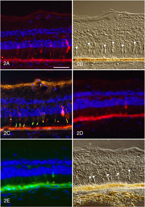
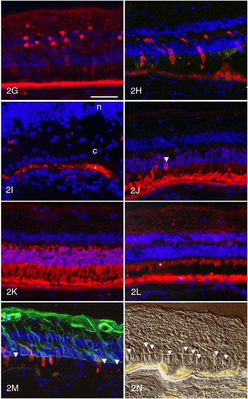
Figure 2. Immunocytochemistry of retina with T-17-M RHO mutation
A. 7G6 labels some cone outer and inner segments, cell bodies and synapses. The remainder of the cones have lost 7G6 immunoreactivity in their inner segments, cell bodies and synapses. B. DIC microscopy of the same field shown in A, illustrating the near normal cytologic appearance of the cones (arrowheads). C. Only a few cones are labeled with 7G6 (red) but most cone outer segments are labeled with anti-red/green cone opsin (green). The cone (*) with a 7G6-positive but red/green cone opsin-negative outer segment is presumably a blue cone. D. Labeling with anti-X-arrestin (red) shows that many cones have lost reactivity for this protein. E. Double labeling of the field in D with 7G6 (green) illustrates that the same cones have lost immunoreactivity for both arrestin and 7G6. F. Nomarski image of field shown in D and E. The cones are indicated by arrowheads. G. Labeling with anti-calbindin illustrates loss of reactivity for this protein in most cones, although the horizontal, bipolar and amacrine cells continue to show strong labeling with the antibody. H. Immunolabeling with anti-IRBP reveals very weak reactivity for this protein (green) in the interphotoreceptor matrix. The cones are labeled (red) with 7G6. I. In the macula, the cone matrix sheaths (red, *) are very short but strongly PNA-positive. c, monolayer of cone nuclei; n, inner nuclear layer. J. In the periphery, the rod outer segments are shortened but show normal intensity of labeling with anti-rhodopsin. One rod (arrowhead) shows cell body labeling with anti-rhodopsin. K. Rod arrestin is localized in the rod outer and inner segments, cell bodies and synapses. L. The rod and cone inner segments (*) are well labeled for the mitochondrial protein, cytochrome C oxidase. M. GFAP labeling is increased in Müller processes in an area where some cones have lost 7G6 reactivity and have ectopic nuclei (arrowheads) in their inner segments. N. DIC of field in M illustrating the cytologic features of the cones (arrowheads).
Autofluorescent lipofuscin is present in the retinal pigment epithelium at the bottom of the panels. The scale bar represents 50 mm.