![]() Figure 4 of
Poehner, Mol Vis 2000;
6:192-198.
Figure 4 of
Poehner, Mol Vis 2000;
6:192-198.
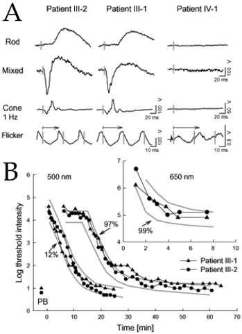
Figure 4. Visual function tests in the South American family
Visual function in the heterozygous parents and homozygous affected daughter from pedigree of Figure 3. A. Rod, mixed cone-rod, and cone electroretinograms in the heterozygous parents (left and middle columns) and in the homozygote (right column). A special protocol was used to record small signals in response to flicker (29 Hz) stimulation in the patient. Vertical bars indicate stimulus onset; calibrations are below and to the right of responses. B. Dark adaptometry results at 30 degrees temporal field in the heterozygous parents after partial (12% rhodopsin) and full (97% rhodopsin, 99% of L/M cone pigment) bleach tests with a 500 nm stimulus. The first 8 min following the full bleach (inset) was tested with the 650 nm stimulus to analyze the L/M-cone recovery. The pre-bleached baseline thresholds are marked with "PB". Connected symbols are the data from heterozygotes; gray lines delimit range of normal results.