![]() Figure 2 of
Poehner, Mol Vis 2000;
6:192-198.
Figure 2 of
Poehner, Mol Vis 2000;
6:192-198.
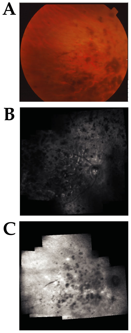
Figure 2. Sardinian family
A. Color fundus photograph of the right eye of patient III-2. Pigmentary disturbances were in the form of clumps or bone spicule-like lesions in the periphery and throughout the posterior pole. A wide area of chorioretinal atrophy included the macula and the peripapillary area. B. Green light reflectance image (mosaic) of right fundus of patient III-3. Pigmentary lesions are distributed throughout the posterior pole and periphery. The atrophy of retinal pigment epithelium at the posterior pole and around the optic disc is severe. C. Infrared fundus image (mosaic) of right eye of patient III-2. The distribution of pigment throughout the posterior pole is again evident.