![]() Figure 5 of
Wong, Mol Vis 2000;
6:184-191.
Figure 5 of
Wong, Mol Vis 2000;
6:184-191.
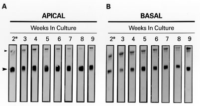
Figure 5. Analysis of apical and basal clusterin from polarized monkey RPE cells in culture
RPE cells were cultivated on filter inserts as described in Methods. Shown are photographs of the immunostained blots of conditioned media samples taken at weekly intervals starting after the initial two weeks in culture. Equivalent volumes of apical (A) and basal (B) medium per sampling were resolved by 12.5% SDS-PAGE and subsequent western analysis using the G7 anti-clusterin antibody at a dilution of 1:1. Two subsets of bands are detected, a set that represents high molecular weight clusterin species (HMW, >100 kDa), and an 80 kDa clusterin species (*80 kDa). Densitometric and statistical analysis of the clusterin immunoreactivity indicate that the clusterin secretion profiles do not change over time. Total clusterin secretion favors a slightly basal profile (1:2 apical:basal), the *80 kDa species considered alone is more equally bi-directionally secreted (1:1.5 apical:basal), and the HMW species is predominantly secreted (1:3.3 apical:basal). Small arrow head: HMW clusterin species. Large arrow head: *80 kDa clusterin species.