![]() Figure 4 of
Wong, Mol Vis 2000;
6:184-191.
Figure 4 of
Wong, Mol Vis 2000;
6:184-191.
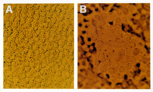
Figure 4. Morphology of polarized monkey RPE cells in culture
Panel A is a photomicrograph of first passage monkey RPE cells in a confluent monolayer culture using pseudo-interference optics. (Magnification: 350x). Panel B is a phase contrast photomicrograph of monkey retinal pigmented epithelial cells at confluence in first passage culture. RPE cells comprising the apex of a fluid-filled dome are in focus, with surrounding cells attached to the plastic culture dish beyond the plane of focus. The appearance of domes in epithelial cultures is emblematic of vectorial ion and small molecule transport, generating hydrostatic pressure from fluid movement to a basal compartment between the monolayer of cells and the impermeable plastic substrate, counterbalanced by the resistance of circumferential tight junctions linking the cells. (Magnification: 350x).
