![]() Figure 3 of
Wong, Mol Vis 2000;
6:184-191.
Figure 3 of
Wong, Mol Vis 2000;
6:184-191.
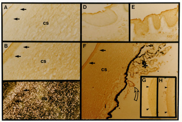
Figure 3. Ocular clusterin localization by immunohistochemistry and in situ analysis
Shown are sections of cornea (panels A-C, serial sections), skin/eyelid (panels D, E, serial sections), ciliary body (unfilled arrow and * in panel F), and lens (panels G, H, serial sections). Immunodetection with G7 are shown in panels B, E, F, and H; panels A, D, and G are the negative controls in which the G7 antibody was omitted. In situ hybridization of an anti-sense clusterin mRNA probe on cornea is shown in panel C. Panel I: graph illustrating the distribution of silver grain over different regions of the cornea shown in panel C, for each general region 10 independent counts within a fixed area were taken. Solid arrows indicate the interface between epithelial and stromal cells (cs) in the cornea, arrow heads indicate the area of epithelial cells in the lens.