![]() Figure 5 of
Seigel, Mol Vis 2000;
6:157-163.
Figure 5 of
Seigel, Mol Vis 2000;
6:157-163.
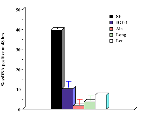
Figure 5. IGF-1 and analogs inhibit apoptosis of retinal cells under serum-free conditions
For serum-free treatment, R28 cells were plated on coverslips and allowed to attach for 6-8 h. After cell attachment, 50 ng/ml IGF-1 or IGF-1 analogs were added and allowed to incubate for the entire 48 h time period. Control coverslips were kept in serum-free medium to promote cell death, as well as serum-containing medium as a negative control. After 48 h, the cells were fixed with either 6:1 methanol: PBS for ssDNA reaction, or in 4% paraformaldehyde for TUNEL-in situ staining. Panel A indicates the percentage of TUNEL positive cells at 48 h for each condition, while panel B demonstrates the percentage of ssDNA-reactive cells. Error bars indicate standard deviations. IGF-1. All analogs showed statistically significant differences from serumfree cultures (p<0.05) except [Leu34][Ala31] in panel A.
A.
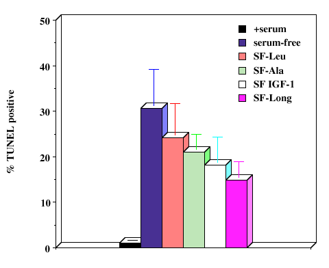
B.