![]() Figure 2 of
Seigel, Mol Vis 2000;
6:157-163.
Figure 2 of
Seigel, Mol Vis 2000;
6:157-163.
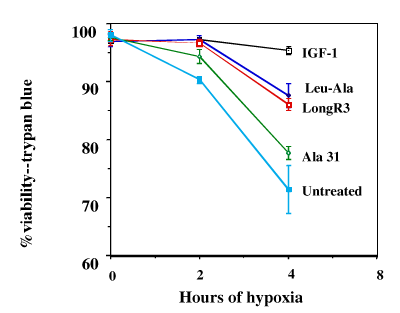
Figure 2. IGF-1 and analogs enhance viability of hypoxic retinal cells
The R28 retinal cell line was subjected to 0, 4, and 8 h of hypoxia (95% N2/5% CO2). IGF-1 and analogs ([LongR3], [Ala31], and [Leu24][Ala31]) were used at a concentration of 50 ng/ml with pre-treatment time of 24 h. Cell survival was determined by trypan blue viability testing. Error bars indicate standard deviations. There were statistically significant differences between untreated cells vs. IGF-1 or analog treatment at 4 and 8 h, with the exception of [Ala31].