![]() Figure 1 of
Yeagle, Mol Vis 2000;
6:125-131.
Figure 1 of
Yeagle, Mol Vis 2000;
6:125-131.
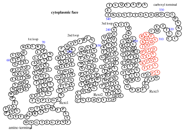
Figure 1. Schematic diagram of the primary sequence of bovine rhodopsin
This figure represents the portions of the polypeptide chain [16] that are now known, from this and other work, to be in an a-helical conformation. The red region indicates the sequence from rhodopsin represented in rhoviih whose structure is described in this report.