![]() Figure 2 of
Ablonczy, Mol Vis 2000;
6:109-115.
Figure 2 of
Ablonczy, Mol Vis 2000;
6:109-115.
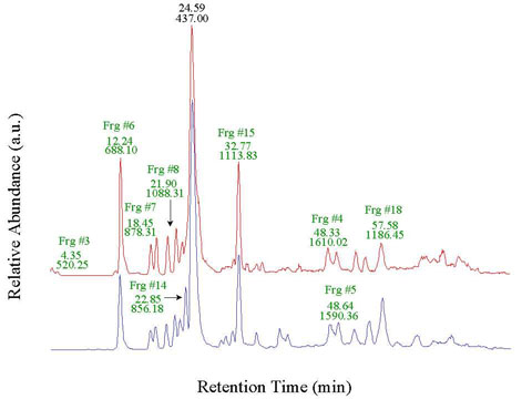
Figure 2. LCQ base peak chromatograms of rat rhodopsin
The chromatograms of dark reared light damaged rhodopsin (top) and the cyclic dim light reared control rhodopsin (bottom) are shown. The most abundant CNBr fragments are indicated. Other fragments can also be found in the background. Additional peaks are contaminants. The retention time (numbers on top) and the mass of the most intense precursor ions (numbers below) are shown. There were no sequence differences or other modification observed between control and light damaged, dark and cyclic dim light reared animals.