![]() Figure 4 of
Ying, Mol Vis 2000;
6:101-108.
Figure 4 of
Ying, Mol Vis 2000;
6:101-108.
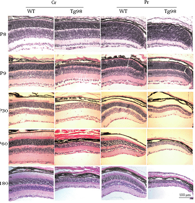
Figure 4. Retina sections from the Tg98 mouse and a wild-type littermate during early postnatal development
At P8, no obvious morphological differences appeared between Tg98 mouse and WT mouse. At P9, in the central retina, the ONL of Tg98 mouse was thinner than that of WT mouse. There was no obvious difference in the peripheral retina between Tg98 and WT. At P30, in the central retina, one single photoreceptor cell layer remained in the ONL of Tg98 mouse. In the peripheral retina, 2 to 3 layers of photoreceptors remained in the ONL of Tg98 mouse. At P60, the entire ONL in the central retina of Tg98 mouse had disappeared. One single layer of photoreceptors remained in the peripheral retina of Tg98 mouse. At P180, in the inferior retina, the entire ONL was absent in both central and peripheral retina of Tg98 mouse. RPE, retina pigment epithelium; OS, outer segment; ONL, outer nuclear layer; INL, inner nuclear layer; G, ganglion cell layer; Cr, central retina; Pr, peripheral retina. H& E-staining. The bar in the lower right panel represents 100 mm.