![]() Figure 2 of
Fujii, Mol Vis 2000;
6:1-5.
Figure 2 of
Fujii, Mol Vis 2000;
6:1-5.
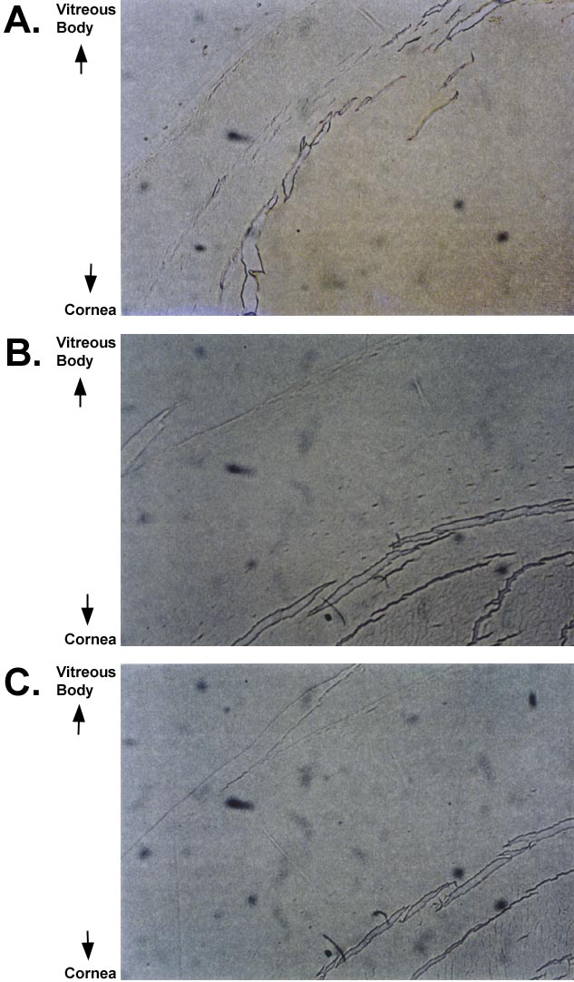
Figure 2. Immunohistochemistry with purified peptide 3R antibody
A shows a 60-year-old eye with significant staining, especially in the core of the lens. B shows negative staining of the 2-year-old eye. C shows control for nonspecific secondary binding without peptide 3R antibody. All photographs were taken using a 50x objective.