![]() Figure 3 of
Duncan, Mol Vis 1999;
5:9.
Figure 3 of
Duncan, Mol Vis 1999;
5:9.
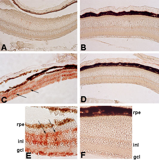
Figure 3. Immunohistochemical detection of retinaldehyde dehydrogenase (AHD2) during postnatal development of Mitfvit and C57BL/6 control mice eyes
Whole eyes from Mitfvit and C57BL/6 control mice at 2, 4, and 10 weeks of age were prepared for immunohistochemical analysis of AHD2 as described in Methods. The sections shown here are representative of the central portion of the retina. There was no difference in the level of AHD2 detected between Mitfvit and control mice at 2 (data not shown) and 4 weeks of age (A and B, respectively). At 10 weeks of age a marked increase in retinaldehyde dehydrogenase immunoreactivity was noted in the dorsal neural retina of Mitfvit (C, arrows) mice compared to controls (D). Higher magnification of the Mitfvit dorsal neural retina at 10 weeks (E) shows intense labelling in vertical arrays (arrows). No staining for AHD2 was evident in the central neural retina of control mice at 10 weeks of age (D), even at a higher magnification (F). (Magnification A-D x200; E and F x400).