![]() Figure 2 of
Duncan, Mol Vis 1999;
5:9.
Figure 2 of
Duncan, Mol Vis 1999;
5:9.
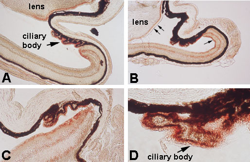
Figure 2. Immunohistochemical detection of retinaldehyde dehydrogenase (AHD2) in postnatal Mitfvit and C57BL/6 control mice ocular tissues
Eyes from age-matched Mitfvit and C57BL/6 control mice were enucleated at 2, 4, and 10 weeks of age. Eyes were prepared for immunohistochemical analysis of AHD2 as described in Methods. Representative sections from C57BL/6 control mice eyes at 2 (A) and 10 (B) weeks of age show typical staining for AHD2. AHD2 was consistently detected and limited to three ocular tissues: the epithelium of the ciliary body, a single layer of cells on the anterior surface of lens, and the far periphery of the dorsal neural retina. The arrows in (B), upper left, point to the anterior portion of the lens that stained positive for AHD2; the single arrow, right side of (B), points to the peripheral portion of the dorsal neural retina that stained positve for AHD2. Representative sections from Mitfvit mice at 4 (C) and 10 (D) weeks of age show the same pattern of staining as controls.