![]() Figure 1 of
Duncan, Mol Vis 1999;
5:9.
Figure 1 of
Duncan, Mol Vis 1999;
5:9.
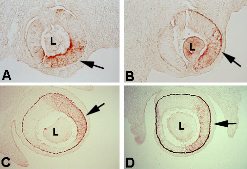
Figure 1. Immunohistochemical detection of retinaldehyde dehydrogenase (AHD2) during embryonic development of Mitfvit and C57BL/6 control mice eyes
Embryos from Mitfvit and C57BL/6 control mice were obtained at E11.5, E13.5 and E15.5. Embryos were prepared for immunohistochemical analysis of AHD2 as described in Methods. AHD2 was detected in the eyes, specifically the dorsal neural retina (arrows), of both Mitfvit and control mice at E11.5 (A and B, respectively), E13.5 (C and D, respectively) and E15 (data not shown). These data show no differences in the staining pattern for AHD2 between Mitfvit and control mice.