![]() Figure 1 of
Haque, Mol Vis 1999;
5:8.
Figure 1 of
Haque, Mol Vis 1999;
5:8.
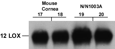
Figure 1. Immunodetection of 12-LOX
A. Electrophoresis of cellular proteins and immunostaining were carried out as outlined in Materials and Methods. Twenty µg of protein from each cell line was loaded in duplicate wells. Lanes 1 and 2, mouse lens epithelial cell line 21EM15; lanes 3 and 4, mouse lens epithelial cell line 17EM15; lanes 5 and 6, rabbit lens epithelial cell line LEP2; lanes 7 and 8, rabbit lens epithelial cell line B3; lanes 9 and 10, human lens epithelial cell line HLE-B3; lanes 11 and 12, mouse lens epithelial cell line [alpha]TN4; lanes 13 and 14, mouse lens epithelial cell line MLE6; and lanes 15 and 16, rabbit lens epithelial cell line N/N1003A.

B. Electrophoresis of cellular proteins and immunostaining were carried out as outlined in Materials and Methods. Twenty µg of protein from each cell line was loaded in duplicate wells. Lanes 17 and 18, mouse cornea and lanes 19 and 20, rabbit lens epithelial cell line N/N1003A.