![]() Figure 3 of
Kuszak, Mol Vis 1999;
5:7.
Figure 3 of
Kuszak, Mol Vis 1999;
5:7.
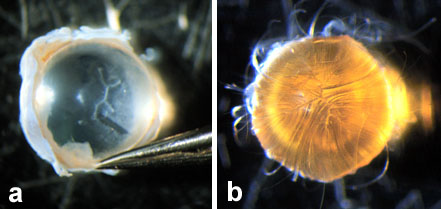
Figure 3. Low power images of RCS rat lenses as seen under a stereo-view dissecting microscope
A representative unfixed 3.5 month old RCS rat lens (A) and a fixed 15 month old RCS rat lens (B), dissected to the axial dimensions of a 6 month old RCS rat lens (intra and inter lens analysis). It is apparent that in both lenses the PSC was internalized by the overlaying of posterior segments of fibers to form abnormal posterior suture patterns. At 3.5 months of age, the resulting posterior suture pattern was an inordinately small, centrally located, inverted Y suture with three secondary branches extending to confluence at the peripheral ends of two of the primary or main suture branches. At 6 months of age, the abnormal posterior suture pattern was slightly more irregular and complex. This suture had an inordinately small, centrally located, inverted Y suture, with three secondary branches extending to confluence at the peripheral ends of two of the main suture branches and two tertiary sub-branches extending to confluence at the peripheral end of one of the secondary branches.