![]() Figure 2 of
Kuszak, Mol Vis 1999;
5:7.
Figure 2 of
Kuszak, Mol Vis 1999;
5:7.
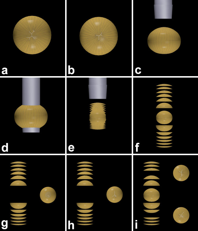
Figure 2. Scale 3D-CADS demonstrating highlights of the dissection protocol used to retrieve complete suture patterns formed in lenses at different ages
(A) The posterior surface of the lens showing an abnormal 9 branch posterior suture pattern. (B) The anterior surface of the lens showing an abnormal 5 branch anterior suture pattern. (C-E) A 3 mm stainless steel trephine is used to remove a cylindrical core from the lens. In this manner, the lateral segments of fibers not involved in suture formation are separated from the anterior and posterior segments of fibers that overlap to form suture branches within and between growth shells. (F) The successive anterior and posterior convexo-concave discs contain, respectively, complete anterior and posterior suture patterns as a function of age overlain onto the PSC (G) and slightly abnormal anterior suture (H) formed from 2-6 weeks after birth. (I) The posterior (upper) and anterior (lower) convexo-concave discs with axial dimensions equal to a 6 month old lens. These discs featured less abnormal posterior and anterior suture patterns than seen in successive discs with axial dimensions equivalent to 9-15 month (A,B) old lenses.