![]() Figure 11 of
Kuszak, Mol Vis 1999;
5:7.
Figure 11 of
Kuszak, Mol Vis 1999;
5:7.
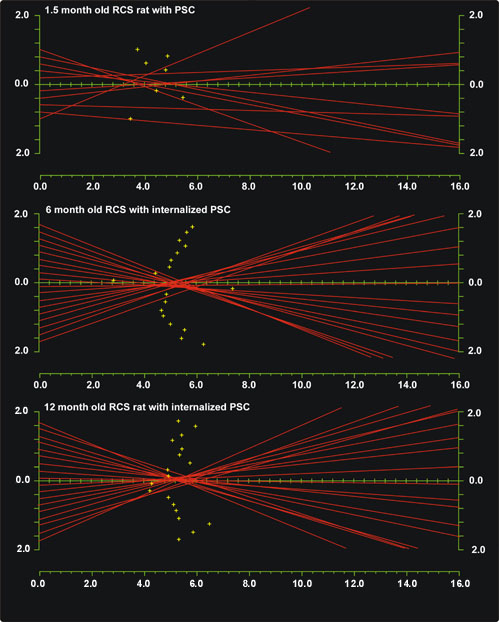
Figure 11. Plots of laser scan analysis through RCS rat lenses
Consider the standard map of laser penetration points directed into a rat lens by our low power helium-neon laser scan analysis as shown previously in Figure 1B. If the lens is turned 90° on its equatorial axis, the variable focal lengths of the eighteen laser beams can be represented as they are reflected or refracted by the lens. The vertical axis indicates laser beam position (mm) from the optical center (0, 0) of the lens. The horizontal axis indicates equivalent focal length (mm). Plus (+) signs indicate the focal lengths for each incident laser beam as a function of distance from the optical center.
RCS rats at 1.5, 6, and 12 month old had a fully formed PSC, a PSC with 4.5 months of internalization by abnormal fiber growth, and a PSC with 10.5 months of internalization by abnormal fiber growth, respectively. Note that while our laser scan analysis routinely directs a laser beams at eighteen locations on a lens per pass (vertical axis), the PSC plaque in 1.5 months old RCS lenses internally reflected almost half of the beams on average. In contrast, internalization of the PSC plaque by abnormal fiber growth effectively produced a larger lens with an added posterior component. This new posterior component allowed the lens to recover the ability to refract all eighteen laser beams.