![]() Figure 10 of
Kuszak, Mol Vis 1999;
5:7.
Figure 10 of
Kuszak, Mol Vis 1999;
5:7.
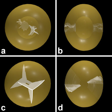
Figure 10. Scale 3D-CADs comparing the abnormal anterior and posterior suture planes formed during internalization of PSC and the normal anterior and posterior suture planes formed during lens growth
In a 15 month old RCS rat lens viewed at a slight angle to the optical axis (A) and along the equatorial plane (B) it can be seen that the complex and abnormal set of suture planes do not extend into the lens periphery. In contrast, in a 15 month old control rat lens viewed at a slight angle to the optical axis (C) and along the equatorial plane (D) it can be seen that the simple normal suture planes of age-matched controls extended farther into the lens periphery.