![]() Figure 8 of
Al-Ghoul, Mol Vis 1999;
5:6.
Figure 8 of
Al-Ghoul, Mol Vis 1999;
5:6.
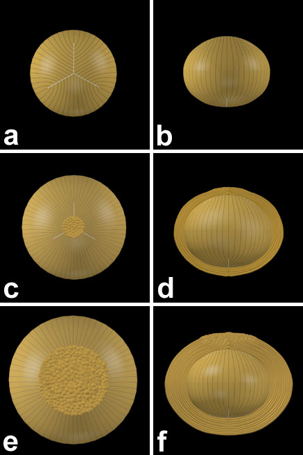
Figure 8. Scale 3D-CAD reconstructions of PSC development in RCS rats
Posterior subcapsular cataractogenesis occurs between 2 and 6 weeks postnatal. At 4 weeks, most lenses had a small, polar opacity which enlarged radially to a large PSC plaque by 6 weeks. The plaque was formed by posterior fiber ends which curved abnormally toward the vitreous and globulized under the capsule. PSC formation precluded development of normal posterior sutures. A posterior view is shown in (A,C,E). An axial view is shown in (B,D,F).
(A,B) 2 weeks postnatal. (C,D) 4 weeks postnatal. (E,F) 6 weeks postnatal.