![]() Figure 7 of
Al-Ghoul, Mol Vis 1999;
5:6.
Figure 7 of
Al-Ghoul, Mol Vis 1999;
5:6.
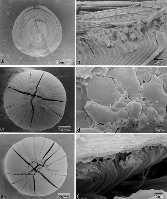
Figure 7. Scanning electron micrographs of internalized plaques at 4, 9, and 15 months old.
The PSC plaques displayed consistent structural features at all ages examined. Specifically, the plaques were composed of markedly enlarged and irregular posterior fiber ends aberrantly curved toward the capsule. (A) 4 month old lens split along the optic axis. (B) Enlargement of boxed area in (A). (C) 9 month old lens dissected to expose the PSC. (D) Enlargement of boxed area in (C). (E) 15 month old lens with the PSC partially exposed. (F) Enlargement of boxed area in (E).
