![]() Figure 3 of
Al-Ghoul, Mol Vis 1999;
5:6.
Figure 3 of
Al-Ghoul, Mol Vis 1999;
5:6.
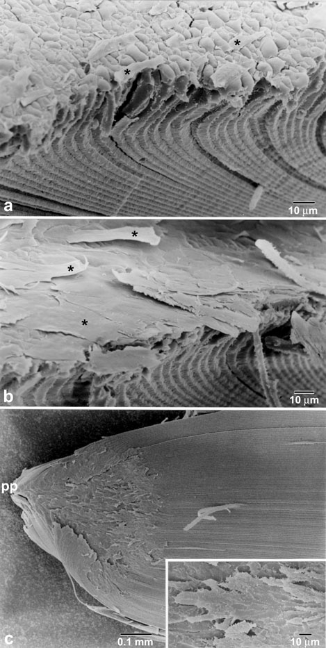
Figure 3. Scanning electron micrographs depicting the morphology of the initial fibers overlying the plaque in 2.5-3 month old lenses.
(A,B) The posterior ends of fibers overlying the plaque were extremely flattened (asterisks). (C) The posterior portion of a fiber peel obtained from dissection of a 3 month old RCS rat lens. Even at low magnification it was apparent that fiber morphology was normal along fiber length but not over the plaque. During internalization of the PSC, each successive shell of fibers terminated progressively closer to the posterior pole (PP), resulting in peripheral to central overgrowth of the plaque. Higher magnification (inset) demonstrating the irregular, branched, terminal extensions of fiber ends overlying the plaque.