![]() Figure 2 of
Al-Ghoul, Mol Vis 1999;
5:6.
Figure 2 of
Al-Ghoul, Mol Vis 1999;
5:6.
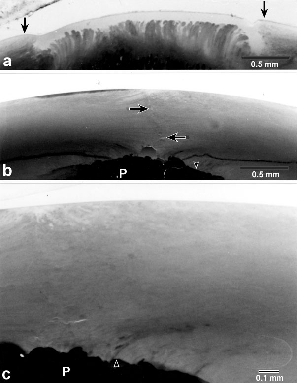
Figure 2. Light micrographs of thick axial sections at the posterior pole during the initial stages PSC internalization.
(A) At 2 months postnatal new fiber growth had accumulated at the periphery of the plaque (arrows) but had not yet overgrown the PSC. (B,C) Non-adjacent serial sections from a 3 month old RCS rat lens. The plaque (P) was completely internalized by new fiber growth. The suture plane formed subsequent to plaque overgrowth is demarcated by arrows (B). At higher magnification (C) it can be seen that overgrowth of the plaque began marginally and advanced to confluence at the pole. The posterior ends of new fibers interfaced with the upturned ends of fibers in the plaque, forming a convexo-concave, disk-shaped suture plane parallel to the capsule (arrowheads).