![]() Figure 10 of
Al-Ghoul, Mol Vis 1999;
5:6.
Figure 10 of
Al-Ghoul, Mol Vis 1999;
5:6.
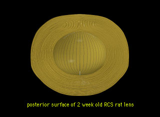
Figure 10. Scale 3D-CAD animation depicting PSC formation and initial PSC internalization in RCS rat lenses
The PSC, which develops between 2 and 6 weeks postnatal, results from a growth malformation of the posterior segments in each successive growth shell, and results in central-to-peripheral plaque formation. Similarly, the plaque is gradually internalized as each growth shell of fibers terminates closer to the posterior pole. This peripheral-to-central overgrowth occurs between 2 and 2.5 months postnatal and results in the formation of an aberrant suture plane across the visual axis.
Note that the slide bar at the bottom of the quicktime movie can be used to manually control the flow of the movie. If you do not want to or are unable to view the movie, a representative frame is included below as a still image.